از همه چیز و همه جا

ما قصد داریم در این وبلاگ مطالب جالب همه رشته ها را قرار دهیم تا شما نیز از آن لظّت ببرید.

از همه چیز و همه جا

ما قصد داریم در این وبلاگ مطالب جالب همه رشته ها را قرار دهیم تا شما نیز از آن لظّت ببرید.

از همه چیز و همه جا
پیام های کوتاه

۰ نظر موافقین ۰ مخالفین ۰ ۱۳ بهمن ۹۳ ، ۱۵:۴۱
M.Y

Abstract

Recombinant protein overexpression of large proteins in bacteria often results in insoluble and misfolded proteins directed to inclusion bodies. We report the application of shear stress in micrometer-wide, thin fluid films to refold boiled hen egg white lysozyme, recombinant hen egg white lysozyme, and recombinant caveolin-1. Furthermore, the approach allowed refolding of a much larger protein, cAMP-dependent protein kinase A (PKA). The reported methods require only minutes, which is more than 100 times faster than conventional overnight dialysis. This rapid refolding technique could significantly shorten times, lower costs, and reduce waste streams associated with protein expression for a wide range of industrial and research applications.

Overexpressed recombinant proteins for industrial, pharmaceutical, environmental, and agricultural applications annually represent a more than $160 billion world market.1 Protein expression in yeast or Escherichia coli is highly preferred, due to the organism's rapid growth, low consumable costs, and high yields.2, 3 However, large proteins overexpressed in bacteria often form aggregates and inclusion bodies.47Recovery of the correctly folded protein then requires laborious and expensive processing of inclusion bodies by conventional methods.1, 8 The most common method for refolding such proteins, for example, involves multi-day dialysis with large volumes (typically 1–10 liters for mg quantities of protein).9 Alternatively, expression of high-value proteins (e.g., therapeutic antibodies or GPCRs for structural biology) require extensively optimized mammalian or insect cell lines, media, and bioreactor conditions.1012 Recovery of correctly folded proteins from aggregates is inefficient and challenging for large-scale industrial processes. Mechanical methods to solve this problem have been reported. One approach applies very high hydrostatic pressures (400 bar) to refold recombinant proteins from inclusion bodies.1316We report a novel method by applying finely controlled levels of shear stress to refold proteins trapped in inclusion bodies. This method might be capable of broadening the utility of bacterial overexpression, and could transform industrial and research production of proteins.

We report using a vortex fluid device (VFD) to apply shear forces for rapid equilibration of protein folding and isolation of intermediates during protein folding. In this method, a glass cylinder (10 mm by 16 cm) is spun rapidly (5 krpm) at a 45° angle. At high rotational speeds, the solution within the sample tube forms micrometer-thick, thin fluid films, which flow with the same speed and direction as the wall of the glass tube. The rotating glass tube generates a velocity gradient within the thin fluid film, which introduces shear stress into the solution (Figure 1 A). We imagined applying the VFD with a similar range of input energies to the refolding of proteins: the device produces unusual shear within such films and has been shown to be effective in disassembling molecular capsules15 and reducing the size of micelles used in the fabrication of SBA-15 mesoporous silica with control over the pore size of the material.16 Importantly, the latter occurs at room temperature rather than requiring hydrothermal processing and suggests that the constant “soft” energy in the thin film has application in manipulating macromolecules, such as the refolding of proteins, without requiring heating. Other relevant applications and optimization of a similar VFD include exfoliating graphite and hexagonal boron nitride to generate mono- and multilayer structures,17, 18 controlling the formation of different calcium carbonate polymorphs,19 and controlling chemical reactivity and selectivity.20, 21

thumbnail image

Figure 1. Protein refolding in vitro with the vortex fluid device (VFD), which generates shear flow inside thin fluid films (shaded). a) VFD schematic and parameters for modeling. The inset represents a two-dimensional slice of the tube through the axis of rotation. The inner diameter of the tube, R2, is 5 mm, and the thin fluid film thickness, δ, has been previously measured (not depicted to scale).17 The velocity of the solution, vθ, increases from the inner surface of the film to match the velocity of the tube, Ω. b) Shear stress calculated as a function of rotational speed (rpm) and radius, R1, from the center of the tube.

Modeling the fluid behavior in the VFD allowed estimation of the shear forces experienced by proteins folding at various rotational speeds. Our analysis applies the solution for cylindrical Couette flow.20, 21 The velocity of the solution, vθ, is a function of the radius, r (Figure 1 A). The boundary conditions for the liquid film interfaces are defined as follows. The inner air–liquid interface at r=R1 slips, due to discontinuity in viscosity, and results in increasingly low shear stress (dvθ/dr=0). At the outer liquid–glass interface, the no-slip boundary dictates that the velocity of the liquid at r=R2 matches that of the inner wall of the glass tube (vθ=R2⋅Ω), where Ω is the angular velocity of the tube. The resulting velocity profile is a nonlinear function of the form(1)

  • equation image
    (1)

where(2)

  • equation image
    (2)

From this velocity profile, shear stress can be calculated as(3)

  • equation image
    (3)

where μ is the viscosity of water at 20 °C. At a speed of 5 krpm, the calculated shear stress ranges from 0.5246 to 0.5574 Pa (Figure 1 B). Although this model provides insight into the shear stress range, the fluid flow will be perturbed by the cross vector of gravity with centrifugal force for a tube oriented at 45°,20 which might also facilitate protein folding; this angular setting also proved optimal for the aforementioned applications of the VFD. The calculated values of shear stress are similar to the levels previously reported to cause protein unfolding,22 and we hypothesized that short periods of VFD processing could allow equilibration of protein folding.

Experiments with native hen egg white were conducted to determine if shear forces could refold denatured hen egg white lysozyme (HEWL) in complex environments. The separated whites were diluted in PBS and heat-treated at 90 °C for 20 min. The resultant hard-boiled egg white was dissolved in 8 m urea, rapidly diluted, and then VFD-processed at the indicated rotational speeds and times (Table S1 and Figure 2 A). The total protein concentration, as determined by bicinchoninic acid assay, was 44 μg mL−1. The recovery of HEWL activity was then demonstrated by a lysozyme activity assay (Figure S1). Refolding HEWL within the egg white at 5 krpm recovered glycosidase activity even after a short 2.5 min spin, but continued shear forces unfolded the protein. As mentioned above, shear-stress-mediated protein unfolding has been observed for other proteins previously. Optimization of VFD times, speeds, and concentration of chaotropic additives is required to find conditions allowing re-equilibration of protein folding without causing loss of the protein structure. VFD processing for 5 min at 5 krpm in 1 m urea resulted in optimal HEWL refolding as monitored by lysozyme activity (Figure 2 A). This experiment establishes the basic parameters for protein refolding by VFD.

thumbnail image

Figure 2. Determination of secondary structure and activity of hen egg white lysozyme (HEWL) processed by VFD. A) Lysozyme activity per mg protein following VFD processing of boiled egg white (90 °C, 20 min) at a fixed VFD speed of 5 krpm or a fixed 5 min refolding time. The relative circle size represents the lysozyme activity, with larger circles representing higher recovered activities, plotted as a function of VFD time and speed. B) CD spectra of recombinantly expressed, reduced, and denatured HEWL before (⋅⋅⋅⋅) and after () VFD refolding; : native HEWL. C) In continuous-flow mode, the protein solution is introduced through a thin, hollow metal tube to the bottom of the sample tube, and the folded protein can be collected at the top of the VFD. D) Lysozyme activity per mg protein following VFD refolding of recombinantly expressed HEWL and native HEWL. Under these conditions, VFD treatment of recombinant lysozyme recovered over 82 % of activity in both confined and continuous-flow modes, compared to wild-type protein (WT). VFD treatment of wild-type, active lysozyme isolated from eggs does not adversely affect its activity (white). Throughout this report, error bars indicate standard deviation (n=3).

To demonstrate refolding of recombinantly expressed, reduced HEWL, the cell pellet was first reconstituted in lysis buffer containing 2-mercaptoethanol, denatured in 8 m urea, and rapidly diluted into PBS (1:100). Buffers used for the expression and purifications of all recombinant proteins are described in Table S2. Second, the diluted protein (1 mL, 44 μg mL−1 protein) was immediately transferred to the VFD sample tube and spun at 22 °C and 5 krpm for 5 min. Circular dichroism (CD) spectra of the VFD-refolded, recombinant HEWL demonstrated restoration of secondary structure from proteins isolated from inclusion bodies. After VFD processing, the CD spectra of identical HEWL samples demonstrated partial recovery of secondary structure compared to the native lysozyme (Figure 2 B). Yields of functional protein determined by lysozyme activity assay are shown in Figure 2 D.

HEWL could also be refolded by continuous-flow VFD. This approach delivers additional sample through an inlet at the cylinder base. The sample (50 mL), added at a flow-rate of 0.1 mL min−1, demonstrated significant recovery of HEWL activity for scalable, high volume applications (Figure 2 C). The recombinant HEWL recovered >82 % of its activity following VFD treatment. As expected, HEWL isolated from inclusion bodies without VFD processing failed to show any lysozyme activity (Figure 2 D). The continuous-flow approach could be readily scaled up and parallelized for industrial applications requiring treatment of very large volumes.

After refolding denatured lysozyme in both complex (egg white) and simple (purified recombinant protein) environments, the next experiments focused on refolding the protein caveolin-1, as an example of a protein requiring an inordinate amount of processing time by conventional approaches (e.g., four days of dialysis). A caveolin variant without its transmembrane domain (caveolin-ΔTM) was recombinantly expressed, and the inclusion body was purified under denaturing conditions. Purified caveolin-ΔTM was diluted and then underwent a short (1 h) dialysis to decrease the urea concentration to 1 m. The protein was then VFD-treated for 0, 10, or 30 min at 5 krpm at a concentration of 186 μg mL−1. The CD spectra of the VFD-processed caveolin-ΔTM showed pronounced minima at 208 and 220 nm, which are indicative of α-helical secondary structure.23 Solution turbidity also decreased sharply following VFD treatment, which illustrates VFD solubilization and refolding of partially aggregated proteins (Figure 3 B). ELISA experiments examined binding by the refolded caveolin-ΔTM to HIV glycoprotein 41 (gp41), a known caveolin binding partner.2426 VFD processing significantly restored protein function, as shown through binding to gp41 (Figure 3 C).

thumbnail image

Figure 3. Determination of secondary structure and activity of caveolin-ΔTM processed by VFD. A) Circular dichroism (CD) spectra of caveolin-ΔTM following VFD-induced refolding or conventional dialysis. B) Turbidity of caveolin-ΔTM, measured by the fractional absorbance at 600 nm compared to the untreated sample, also at 600 nm. The arrow indicates absorbance of caveolin-ΔTM following 4-days of dialysis. Both 30 min of VFD-based refolding and 96 h of dialysis significantly decrease the turbidity of the protein solution. C) Binding of caveolin-ΔTM to gp41 determined by ELISA with untreated, VFD-treated, and conventionally refolded by dialysis. All other conditions and buffers remained identical. Although some binding occurs without VFD treatment, caveolin-ΔTM binds with greater affinity after refolding by VFD. ⋅⋅⋅⋅: untreated, - - - -: 10 min VDF, : 30 min VDF, : 96 h, dialyzed.

Larger-sized proteins initially failed to refold, despite VFD treatment. For example, the catalytic domain of cAMP-dependent protein kinase A (PKA, 42 kDa) is significantly bulkier than HEWL (14 kDa) and caveolin-ΔTM (17 kDa) and did not refold from inclusion bodies after treatment with similar protocols to the above experiments. To refold full-length PKA in vitro, we adapted the VFD to provide a closer mimic of cellular folding. In cells, the nascent polypeptide can fold as the N terminus extrudes from the ribosome, whereas in vitro refolding must address the entire protein at once.27 Thus, we attempted to focus shear stress on the N terminus of His-tagged PKA by binding the unfolded protein to Ni2+-charged immobilized metal affinity chromatography (IMAC) resin. The His6-PKA-IMAC complex (1 mL, 0.2–1 mg mL−1) was then subjected to refolding by VFD. Following VFD treatment, His-PKA separated from the IMAC resin and recovered 69 % of its kinase activity (Figure 4). Interestingly, the remaining His-PKA eluted from the IMAC resin by imidazole yielded far less active protein (Figure S3). We hypothesize that protein folding makes the His6 epitope less accessible for strong binding to the IMAC resin subjected to shear by the VFD. Under conventional conditions, IMAC resin binds to folded PKA, but we recovered protein remaining on the IMAC resin after VFD treatment to demonstrate its lack of folding and catalytic activity (Figure S3). Thus, as the VFD folds the protein, PKA elutes from the affinity resin. Negative control experiments with identical conditions but with uncharged IMAC resin, decreased quantities of charged resin, or 500 mm imidazole to block the Ni2+-His6 tag interaction showed only low levels of kinase activity (Figure 4).

thumbnail image

Figure 4. Facilitating VFD refolding of PKA by pre-binding the His6-tagged protein to IMAC resin. PKA (1.7 mg) was pre-incubated with 250 or 50 μL IMAC resin in 6 m guanidine-HCl prior to dilution to 1 m guanidine-HCl and VFD treatment. Excess PKA was removed by an imidazole (1 mm) wash buffer. The PKA activity per μg of protein was measured by a nicotinamide adenine dinucleotide (NADH) enzyme-linked assay and is shown here as a percentage of untreated, wild-type PKA activity (WT). Dark gray bars denote samples that are VFD-treated. In this assay, the consumption of ATP by PKA catalysis results in a lactate dehydrogenase-coupled decrease in the levels of NADH, monitored through measuring absorbance at 340 nm. The control experiments included low quantities of resin (50 μL IMAC) and two controls for non-specific binding to the resin in the absence of the Ni2+-His6 tag interaction (500 mm imidazole added, and uncharged IMAC resin lacking Ni2+). Imidazole was diluted to 50 mm before the kinase assay.

As reported here, protein refolding by VFD requires optimization for each protein. Buffers, chaotrope additive, protein concentration, and processing time were optimized for HEWL, caveolin-ΔTM, and PKA. The refolding of HEWL from the complex mixture of boiled egg whites appeared less efficient than recovery of the folded protein isolated from inclusion bodies. In egg whites, the mechanical energy of the VFD could be misdirected to the other >96 % of proteins present.

This report offers significant advantages over conventional approaches to protein refolding. First, VFD-mediated refolding requires much smaller solution volumes (ca. 1 % of the volumes required for conventional dialysis). Second, this key step in protein production occurs more than 100 times faster than overnight dialysis, with >1000-fold improvements for proteins such as a caveolin. Notably, introducing high shear in thin fluid films is a low energy, inexpensive process and can be readily implemented for relatively low cost in conventional biochemistry laboratories.

The advantages of VFD refolding open new possibilities for increasing protein yields in simple cell lines. By using the constant soft energy of the thin microfluidic films, the VFD can untangle complex mixtures, aggregates, and insoluble inclusion bodies. In industrial and academic applications, high concentrations of a chemical inducer like IPTG could drive overexpressed proteins into insoluble inclusion bodies before VFD-based refolding. Conversely, most conventional processes avoid inclusion bodies by optimizing growth conditions and special cell lines at the expense of higher yields and purer protein isolated directly from bacterial cells. As reported here, the continuous-flow mode of the VFD or parallel processing with multiple VFD units could allow scale-up to accommodate much larger solution volumes. Thus, the approach could drastically lower the time and financial costs required to refold inactive proteins on an industrial scale. The VFD sample tube itself can also be modified to amplify or otherwise direct the intensity of shear forces applied. For example, modified surfaces with high contact angle and/or with textured features could enhance the turbulent flow, altering the applied shear stress. Harnessing shear forces to achieve rapid equilibration of protein folding could be expanded to a wide range of applications for research and manufacturing.

۰ نظر موافقین ۰ مخالفین ۰ ۱۳ بهمن ۹۳ ، ۱۵:۳۶
M.Y
چگونه می‌توان تخم مرغ پخته را خام کرد؟

پختن یک تخم مرغ شاید ساده‌ترین بخش یک صبحانه یا یک وعده‌ی غذایی باشد(کاری که همه ی دانش جویان به خوبی باید بلد باشند) اما شیمی پنهان شده پشت آن بسیار جالب و جذاب است. در حقیقت 90 درصد یک تخم مرغ از آب تشکیل شده است و 10 درصد باقی مانده آن نیز پروتئین است که ظاهر لزج سفیده نیز از همین پروتئین‌ها ناشی می‌شود. این پروتئین‌ها در حقیقت زنجیره‌های بسیار بلند در هم فرورفته و تاخورده‌ی آمینواسید‌هایی هستند که به وسیله‌ی نیروهای بین مولکولی مشابه رشته‌های ماکارونی در هم تنیده شده کنار هم قرار گرفته‌اند. هنگامی که تخم مرغ را درون آب جوش می‌اندازید حرارت باعث در هم شکستن پیوندهای بین مولکولی (واندروالسی) می‌شود و زنجیره‌ها شروع به تغییر شکل و شکستن می‌کنند. این زنجیره‌ها سپس شروع به برقراری پیوند بین دیگر زنجیره‌های آمینو اسید می‌کنند که نتیجه‌ی آن تبدیل سفیده‌ی تخم مرغ به ماده‌ای ژلاتینی و کاملا سفید می‌شود. پختن طولانی مدت آن نیز باعث پیچیدن زنجیره‌ها به دور خود و بیرون راندن آب و درنتیجه سفت شدن و لاستیکی شدن سفیده خواهد شد.

کاری که دانشمندان در دانشگاه UC Irvine انجام دادند، در حقیقت تعدادی تخم مرغ را به مدت 20 دقیقه در دمای 90 درجه‌ی سانتی گراد پختند تا کاملا سفت شوند. سپس آن‌ها فرایند را به صورت معکوس اجرا کردند و سفیده‌ی سفت شده را با اضافه کردن اوره به پروتئینی شفاف به نام لیزوزوم تبدیل کردند. اوره پیوندهای شیمیایی سفت کننده سفیده را می‌شکند و آن‌ها را از هم باز می‌کند. آن‌ها سپس این ماده‌ی نه چندان دلچسب را به کمک جریان ورتکس و اعمال تنش‌های برشی به سفیده‌ی پیشین قبل از پخت شدن تبدیل کردند. تنش برشی در این جا باعث بازشدن گره خوردگی‌های بین زنجیرها و تبدیل ساختار پروتئین‌ها به ساختار پیش از پخته شدن می‌شود.

البته قرار نیست تخم مرغ بازخام شده به سفره‌های ما راه پیدا کند، اما هدف اصلی این پژوهش بازیابی ساختار مطلوب پروتئین‌ها بود که در روش‌های پیشین تا چهار روز برای چنین کاری زمان نیاز بود اما در اینجا تنها دقایقی برای این کار کافی است. حقیقت این است که ساختارهای پروتئینی بسیاری وجود دارند که استفاده از آن‌ها نیازمند بازگرداندن‌شان به ساختار اولیه دارد.

به گفته‌ی این تیم چنین فرایندی می‌تواند کاربردهای بسیاری در ساخت پروتئین‌های مورد نیاز صنعت و علم داشته باشد، برای مثال دستیابی به آنتی بادی‌های درمان سرطان به روشی ارزان‌تر و سریع‌تر از روش‌های معمول یا حتی پروتئین‌هایی یکسان برای تولید پنیر!

نتایج این پژوهش در نشریه‌ی ChemBioChem به چاپ رسیده است.

شما نیز می‌توانید در صورت علاقه مندی نتایج پژوهش‌های خود را در نشریات معتبر جهانی را با ما و سایر دوستان زومیتی در میان بگذارید.

منبع:zoomit

۰ نظر موافقین ۰ مخالفین ۰ ۱۳ بهمن ۹۳ ، ۱۵:۳۴
M.Y


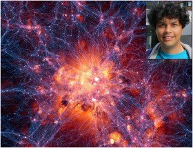
تیمی بین‌المللی از منجمان با همکاری یک محقق ایرانی، شبیه‌سازی‌ای از جهان خلق کرده‌اند که از لحاظ اندازه‌ و سن به جهان مملو از کهکشا‌ن‌های واقعی‌ شباهت دارد.

به گزارش سرویس علمی ایسنا، این موضوع امکان انجام تحقیقات بیشتر درباره چگونگی تکامل‌یافتن جهان به شکل کنونی‌اش طی 14 میلیارد سال را فراهم می‌کند.

این محققان، به رهبری پروفسور «جوب شایه» از دانشگاه لایدن هلند و با همکاری علیرضا رحمتی، محقق ایرانی و دانش‌آموخته دانشگاه‌های شریف، امیرکبیر و لایدن، از ابررایانه‌های دانشگاه دورهام و نیز ابررایانه‌های موجود در پاریس برای خلق شبیه‌سازی موسوم به «تکامل و مونتاژ کهکشان‌ها و محیط‌ها» (EAGLE) استفاده کردند.

تیم علمی توانست انعکاسی دقیق از کهکشان‌های واقعی را با واردکردن بادهای قوی کهکشانی در این شبیه‌سازی وارد کند. این بادها از ستارگان، انفجارات ابرنواختری و سیاهچاله‌های فوق عظیم ناشی می‌شوند و منبع گازی لازم برای شکل‌گیری ستاره‌ها و کهکشان‌هایی سبک‌تر و جوان‌تر را ارائه می‌دهند.

در شبیه‌سازی‌های پیشین، بادهای کهکشانی ضعیف‌تر بودند و منجر به شکل‌گیری زودهنگام کهکشان‌ها در این مدل، در مقایسه با جهان مشاهده‌شده دقیق می‌شد.

به گفته «ریچارد باور» از دانشگاه دورهام، جهان خلق‌شده با استفاده از رایانه در این تحقیق درست مانند جهان واقعی است.

در مدل‌ شبیه‌سازی‌شده، کهکشان‌ها با تمامی شکل‌ها، اندازه‌ها و رنگ‌ همه جا هستند و این درست مانند آن چیزی است که دانشمندان با استفاده از تلسکوپ‌های عظیم مشاهده کرده‌اند. در این جا، حتی می‌توان با فشاردادن دگمه‌ای، زمان را به عقب بازگرداند.

با داشتن مدلی که کهکشان‌ها را درست مانند جهان واقعی تولید می‌کند، محققان امیدوارند بتوانند از آن به عنوان ابزاری برای کنکاش چگونگی به وجودآمدن جهان پس از انفجار بیگ‌بنگ استفاده کرد.

جزئیات این تحقیقات در نشریه Monthly Notices from the Royal Astronomical Society قابل‌ مشاهده است. 

۰ نظر موافقین ۰ مخالفین ۰ ۰۷ بهمن ۹۳ ، ۱۵:۵۱
M.Y

 به نقل از آرت دیلی؛ متخصصان آمریکایی در حال بررسی تفنگی قدیمی هستند؛ این تفنگ که نوعی اسلحه وینچستر مدل 1873 است توسط یکی از کارکنان پارک ملی آمریکا در "نوادا" که در حال همکاری با گروه باستان شناسی حاضر در این پارک بود، پیدا شد. 


قسمت چوبی تفنگ فوق به دلیل سال‌ها مجاورت با هوا و عناصر طبیعی دچار هوازدگی و ترک شده در حالیکه لوله زنگ زده آن با رنگ درخت "ارس" آمیخته شده است. 


این تفنگ به علت گیر کردن در یک بیرون آمدگی سنگی سالها از دید همه پنهان بوده است، شماره سریال این تفنگ نشانگر ساخت آن در سال 1882 است، اما محققان به دنبال اطلاعاتی نظیر مشخصات صاحب آن، دلیل تکیه دادن آن به درخت و یا پنهان ماندنش برای مدت طولانی هستند. 


این نوع اسلحه که مدل 1873 وینچستر است از نظر تاریخی اهمیت زیادی در آمریکا دارد چراکه مهاجران غرب آمریکا از آن در جنگ داخلی این کشور استفاده می‌کرده‌اند و به همین دلیل علت آن را "فاتح غرب" می‌نامند. 


در بین سالهای 1873 تا 1916 حدود 720 هزار‌ و 610 عدد از این نوع اسلحه تولید شده است. 

این تفنگ در زمان اکتشاف خالی بوده ولی در زمان استفاده حدود 40 الی 44 قطار فشنگ از آن شلیک شده است.
۰ نظر موافقین ۰ مخالفین ۱ ۰۷ بهمن ۹۳ ، ۱۵:۵۰
M.Y

سلام بر دوستان عزیز.

پیشنهاد می کنم حتماً به وبلاگ زیر سر بزنید.

www.ghadirioon.blog.ir

۰ نظر موافقین ۰ مخالفین ۰ ۰۲ بهمن ۹۳ ، ۱۵:۱۱
M.Y

درخشندگی کرم شب‌تاب به دلیل حضور اکسیژن و وقوع واکنش اکسید شدگی در اندام‌های این حشره است. دانشمندان توسط تصویربرداری اشعه‌ی ایکس و همچنین میکروسکوپی اندام‌ها با قدرت تفکیک 20 نانومتر به بررسی چگونگی آن پرداخته‌اند.

کرم شب‌تاب برای جفت‌گیری و برقراری ارتباط به تولید نور چشمک‌زن می‌پردازد. این کار توسط یک فرآیند شیمیایی شناخته شده صورت می‌گیرد که در آن در حضور اکسیژن، آنزیمی بر روی ترکیبِ لوسیفرین (Luciferin) عمل می‌کند (لوسیفرین عنوان لاتین "مبشر نور" است). این کار باعث می‌شود واکنش اکسید شدگی رخ دهد که خود با انتشار نور همراه است. تعیین مکانیزمی که در آن اکسیژن به صورت منظم تهیه می‌شود، دشوار است. زیرا شبکه‌ی نای مانندی که گاز را به اندام‌هایی که نور را منتشر می‌کنند )photocites) می‌برد، ابعاد کوچکی از مرتبه‌ی 100 نانومتر دارد. این ابعاد از توان تفکیک اغلب میکروسکوپ‌ها کوچک‌تر است. اکنون ییکانگ هوو و همکارانش در تایوان از روش‌های میکروسکوپی و توموگرافی با قدرت تفکیک 20 نانومتر برای تصویر کردن اندام‌های حشرات و به‌دست آوردن شار اکسیژن حدودی که برای مکانیسم مورد نظر لازم است، استفاده کرده‌اند.

محققان آزمایش‌های خود را به‌گونه‌ای طراحی کرده‌اند که بتوانند توسط آن‌ها دو فرضیه اصلی برای مکانیزم تهیه اکسیژن را محک بزنند. فرضیه‌ی اول حول تولید گاز اکسید نیتریک (NO) درون اندام‌هایی که نور را منتشر می‌کنند، می‌چرخد. قبل از هر درخشش، گاز به درون میتوکندری (اندامکی که توسط اکسیژن تغذیه می‌شود و تولید انرژی درون سلول را کنترل می‌کند) رسوخ کرده و فعالیت آن را متوقف می‌سازد و در نتیجه اکسیژن اضافی برای پدیده‌ی شب‌تابی باقی می‌ماند. فرضیه‌ی دوم اینگونه مطرح می‌شود که مایع در حال گردش در شبکه‌ی نای گونه برای کنترل میزان اکسیژن کم و زیاد می‌گردد. آزمایشات محققان در سینکروتون منبع نور تایوان انجام شده است. این آزمایش‌ها دلیلی برای اثبات تئوری اول است. تصاویر ثبت شده از سیستم نای‌گونه‌ی حشرات زنده که توسط اشعه‌ی ایکس ثبت شده است، هیچ شاری را درون سیستم نای‌گونه نشان نمی‌دهند. محققان قادر هستند توسط بررسی با کیفیت بالای هندسه‌ی سیستم نای‌گونه، میزان گازی که از شبکه‌ی نای‌گونه به سمت بافت‌ها منتشر می‌شود را تخمین بزنند. نتیجه ‌ی این آزمایش‌ها مقادیری است که با فرضیه‌ی اول (گاز اکسید نیتریک) سازگار است. 

۰ نظر موافقین ۰ مخالفین ۰ ۲۴ دی ۹۳ ، ۱۷:۱۶
M.Y

  محققان سنگاپوری با استفاده از نانوذرات طلا، موفق به ساخت پیمایشگری شدند که قادر است سلول‌های سرطانی را شناسایی کند.

به گزارش سرویس علمی ایسنا، پژوهشگران مؤسسه آاستار موفق به ساخت نانوذرات پلیمری هیبریدی شدند که در محیط اسیدی اطراف تومور سرطانی، نور منتشر می‌کند. این نانوپیمایشگر می‌تواند وجود ذرات کوچک تومور سرطانی را در بدن نشان دهد و برای تشخیص زودهنگام بیماری استفاده شود.

سلول‌های سرطانی دارای محیط اسیدی هستند، به طوری که pH درون این سلول‌ها و در محیط اطراف آن‌ها اندکی کمتر از سلول‌های طبیعی است. این تفاوت، دستمایه پروژه‌های مختلفی شده که در آن محققان به دنبال ساخت پیمایشگرهایی برای شناسایی اسیدیته تومورها هستند.

در بیشتر این پیمایشگرها، اسیدیته فضای درون سلول مورد نظر است؛ بنابراین لازم است که پیمایشگر وارد سلول شود. اما مشکل اصلی در اینجا آن است که بتوان تفاوت میان pH سلول‌های سالم و تومور را به‌ دست آورد.

محققان این پروژه می‌گویند: هدف ما این است که روشی برای روشن کردن تومور در تصاویر گرفته شده از بدن ارائه کنیم.

این گروه تحقیقاتی با خودآرایی نانوذرات طلا و نوعی پلیمر، موفق به ساخت پیمایشگری شدند که در سلول‌های سالم هیچ انتشار نوری نداشته، اما با افزایش مقدار اسیدیته سلول و رسیدن به رقم تقریبی 6.5، گروه‌های شیمیایی موجود در آن تغییر کرده و پیمایشگر نشر فلورسانس دارد.

این گروه تحقیقاتی برای اثبات عملکرد این پیمایشگر، از آن برای شناسایی سلول‌های سرطانی موجود در محیط کشت سلولی استفاده کردند و در نهایت نشان دادند که زیر نور سفید، این پیمایشگر می‌تواند محل سلول‌های سرطان پستان موش را نشان دهد.

این گروه، پیمایشگر را به موش‌ها تزریق کرده و بعد از چهار ساعت، به وضوح نشر فلورسانس را در محل تومور مشاهده کردند. از آنجایی که این روش تشخیص، غیرمخرب است، بنابراین می‌توان از آن برای آزمون‌های انسانی استفاده کرد.

این محققان می‌گویند: این نانوپیمایشگر زیست‌سازگار، عاملی بسیار مهم در حوزه پزشکی است. ما قصد داریم اثرات این نانوپیمایشگر را روی بدن مورد بررسی قرار داده تا از غیرسمی بودن آن مطمئن شویم. 

۰ نظر موافقین ۰ مخالفین ۰ ۲۴ دی ۹۳ ، ۱۷:۱۴
M.Y

جهت رعایت عدالت و برابری:

آهای دختر سرزمین من . . .

اخ ببخشید باز  فراموش کردم !

پسر سرزمین من ,

دیروز مردان غیورسرزمینم مردانه در میدان میجنگیدند...

معلوم هست کجایی خیلی وقت است تو را گم کرده ایم !!!

امروز داری توی آرایشگاه ها ابرو بر میداری،مو رنگ میکنی،دماغت را سر بالا میکنی..

اهای پسر سرزمین من . . .

دختران سرزمین من نیاز به مردی دارند که محکم باشد،قوی باشد در سختی ها

در دشواری ها همراه و همیارشان باشد انقدر قوی وغیور باشد ....

که همه دنیا در برابرش کم بیاورد...

اهای پسر سرزمین من...!!!!!!!

۰ نظر موافقین ۰ مخالفین ۰ ۲۰ دی ۹۳ ، ۱۳:۱۴
M.Y


اس ام اس


دختر سرزمین من!


احساس مےکنم این روزها معناے زیبایے را گم کرده اے


باور کن زیبایے در چشمهاے رنگ روشن و لبهاے برجسته کرده سرخ نیست


باور کن هر چه قدر هم دماغت را کوچک و


سر بالایش کنے زیبا نخواهے شد...


معیارهاے زیبایے ات را تغییر بده


زیبایے ظاهرے هر فرد،همانیست که خداوند در وجودش 


قرار داده...


دختر سرزمین من


این زیبایے ظاهر زمانے به چشم خواهد آمد که تو قلب پاکے داشته باشے


آنوقت همه تو را زیبا خواهند دید!!


از صمیم قلب برایت آرزو مےکنم زیبایے حقیقے گم شده ات را دوباره پیدا کنے

۰ نظر موافقین ۰ مخالفین ۰ ۲۰ دی ۹۳ ، ۱۳:۱۲
M.Y